![]() Figure 3 of
Wang, Mol Vis 2006;
12:76-84.
Figure 3 of
Wang, Mol Vis 2006;
12:76-84.
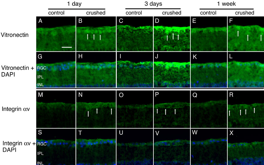
Figure 3. Increased expression of vitronectin and integrin αv in the retina after optic nerve crush
Section of crushed (B,D,F,H,J,L,N,P,R,T,V,X) and contralateral control (A,C,E,G,I,K,M,O,Q,S,U,W) retinas were stained with antivitronectin antiserum (A-L), anti-integrin αv antiserum (M-X), and DAPI (G-L,S-X) at one day, three days, and seven days after optic nerve crush. Induction of vitronectin and integrin αv was found within the retinal ganglion cell (RGC) layer in the crushed group at 1 day to 1 week postcrush (B,D,F,N,P,R, arrows), compared with the control retina. The inner plexiform layer (IPL) and inner nuclear layer (INL) are identified. The scale bar represents 25 μm.