![]() Figure 2 of
Wang, Mol Vis 2006;
12:76-84.
Figure 2 of
Wang, Mol Vis 2006;
12:76-84.
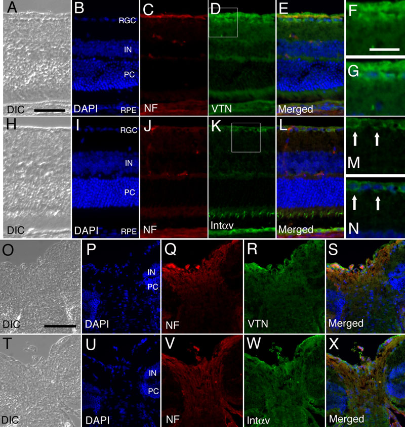
Figure 2. Expression of vitronectin and integrin αv in the retina and the optic nerve head
Sections of normal retina (A-N) and optic nerve head (O-X) were reacted with anti-vitronectin antiserum (VTN; D,F,R, green), anti-integrin αv antiserum (Intαv; K,M,W, green), anti-neurofilament antibody (NF; C,J,Q,V, red) and DAPI (B,I,P,U, blue); merged photomicrographs are also shown (E,G,L,N,S,X). Differential interference contrast light microscopic views are shown in A,H,O,T. Vitronectin is expressed in the nerve fiber layer and retinal ganglion cell (RGC) layer, inner and outer plexiform layer, inner segment of photoreceptor (PC), and retinal pigment epithelium (RPE). The nerve fiber layer is revealed by neurofilament staining. Immunostaining of integrin αv is present in inner retina, inner and outer nuclear layers, outer plexiform layer, inner segment of photoreceptors, and retinal pigment epithelium. The staining pattern of integrin αv in the RGC layer is seen in high magnification images (F,G and M,N from D and K, respectively). The inner nuclear (IN) layer is also identified. In the normal optic nerve head, vitronectin and integrin αv are weakly expressed. Rows of glial cells, indicated by DAPI staining, are found in the septum (between the nerve bundles) of the optic nerve head. The scale bars represent 50 μm in A-E,H-L; 25 μm in F,G,M,N; and 100 μm in O-X.