![]() Figure 6 of
Shirai, Mol Vis 2006;
12:681-691.
Figure 6 of
Shirai, Mol Vis 2006;
12:681-691.
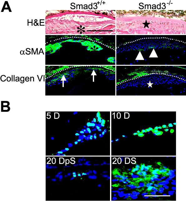
Figure 6.
Development of alkali burn-induced subcapsular cataract is attenuated, but not completely blocked, by loss of Smad3. A: Histology and immunolocalization of αSMA and collagen type VI in Smad3-/- mouse lens at 20 days following alkali injury. Hematoxylin and eosin staining revealed lens epithelial cells are elongated, fibroblast-like in their shape (asterisk) in Smad3+/+ mice, while they are relatively ovoid and epithelial-like in morphology (star) in Smad3-/- mice at day 20 post-alkali exposure. αSMA is markedly expressed in almost all the cells (asterisk) beneath the anterior capsule in a Smad3+/+ lens, whereas faint, but positive, αSMA immunoreactivity is detected in a Smad3-null lens. In a Smad3-null lens αSMA-positive lens epithelial cells are mainly detected on the border between the cell mass and lens cortex (arrowheads). Type VI collagen is detected in the lens of a Smad3+/+ mouse (arrows), but not in a Smad3-/- lens (star). Nuclei were counterstained with DAPI and the scale bar represents 50 μm. B: Immunolocalization of phospho-Smad2 and Smad3 in mouse lens following alkali injury. Phospho-Smad2 is detected in the nuclei of lens epithelial cells at day 5 and 10. The number of positive phospho-Smad2 nuclei seems less at day 20 as compared with earlier times. Smad3 is located in the cytoplasm and nuclei of lens cells in the cell multilayer at day 20. Smad3 and phsopho-Smad2 are not detected in the nuclei of epithelium of an uninjured lens of either Smad3+/+ or Smad3-/- mice (not shown). Nuclei were counterstained with DAPI. The bar represents 20 μm.