![]() Figure 3 of
Shirai, Mol Vis 2006;
12:681-691.
Figure 3 of
Shirai, Mol Vis 2006;
12:681-691.
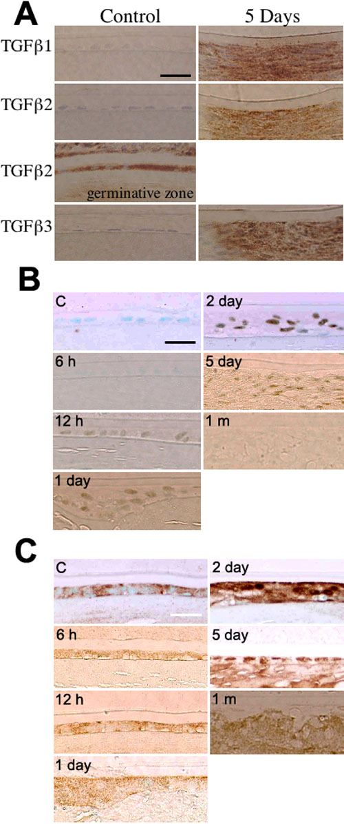
Figure 3.
Transforming growth factor β (TGFβ) expression and its signaling in lens or aqueous humor of an alkali-burned rat eye. A: Immunolocalization of TGFβ1, 2, and 3 in rat lenses at day 5 following alkali injury. TGFβ1 and TGFβ3 are not detected in the uninjured lens and at day 5. TGFβ2 was detected in only the germinative zone of lens epithelial cells in the uninjured lens. At day 5, epithelial mesenchymal transition (EMT) lens cells express TGFβ1, TGFβ2, and TGFβ3. B: Immunolocalization of phospho-Smad2 in rat lenses following alkali injury. At 12 h, phospho-Smad2 protein is weakly detected in the nuclei in the lens epithelial cells and then is detected in the nuclei of the majority of elongated cells in the cell multilayer of the cataractous area through days 1-5. It is no longer detected in such elongated cells in the cataractous region at 1 month. C: Immunolocalization of Smad3 in rat lenses following alkali injury. Smad3 protein is detected in the cytoplasm of uninjured lens epithelial cells at 6 h to day 1. At days 2 and 5, Smad3 protein is detected in the nuclei of fibroblastic cells as well as in their cytoplasm. It is, however, detected in the cytoplasm, but not the nuclei at 1 month. Nuclei were counterstained with methylgreen (A-C). The bars represent 10 μm. D: TGFβ2 levels in injured rat lens and aqueous humor. Active and total TGFβ levels were assayed using ELISA. The asterisk indicates a p<0.05 and the double asterisk indicates a p<0.01; nonsignificant comparisons are marked "NS".