![]() Figure 2 of
Shirai, Mol Vis 2006;
12:681-691.
Figure 2 of
Shirai, Mol Vis 2006;
12:681-691.
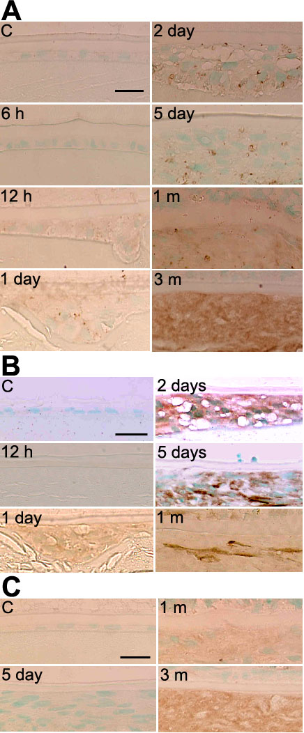
Figure 2.
Expression pattern of epithelial mesenchymal transition (EMT) markers in rat lenses following alkali injury. A: Immunolocalization of Snail in rat lenses following alkali injury ("C" indicates control tissue). Snail protein is detected weakly in lens epithelial cells at 12 h. Its expression increased in EMT lens cells at the later times until 3 months. B: Immunolocalization of αSMA in rat lenses following alkali injury ("C" indicates control tissue). The lens epithelial cells that have formed multiple layers beneath the anterior capsule weakly express αSMA at day 1. At day 2, elongated cells multilayered beneath the anterior lens capsule are markedly labeled with the antibody. C: Immunolocalization of collagen type I in rat lenses following alkali injury ("C" indicates control tissue). Collagen type I is weakly detected in EMT lens cells at one month and strongly positive at three months. Nuclei were counterstained with methylgreen. The bars in the control images represent 10 μm.