![]() Figure 1 of
Shirai, Mol Vis 2006;
12:681-691.
Figure 1 of
Shirai, Mol Vis 2006;
12:681-691.
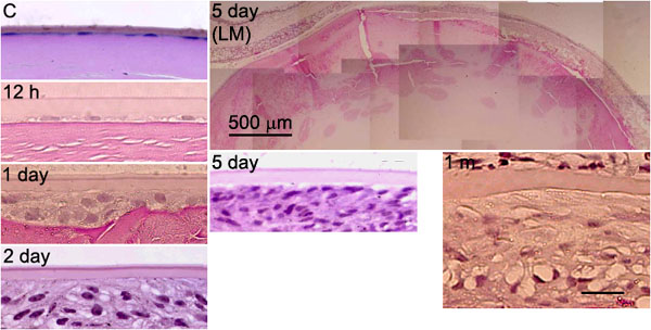
Figure 1.
Alkali injury to the ocular surface induces anterior subcapsular cataract. At 12 h, anterior lens epithelial cells partially disappeared, as compared to the control (C) tissue. At day 1, lens epithelial cells formed multiple layers beneath the anterior capsule. At day 2, the layers of spindle-shaped fibroblastic cells were observed beneath the anterior capsule and became prominent after day 5. At 1 month (1 m), fibrous tissue was observed beneath the anterior capsule. The tissues were stained with hematoxylin and eosin. The bar in the low magnification (LM) 5 day image represents 500 μm; the bar in 1 m represents 10 μm and applies to all other images.