![]() Figure 1 of
Roberts, Mol Vis 2006;
12:633-638.
Figure 1 of
Roberts, Mol Vis 2006;
12:633-638.
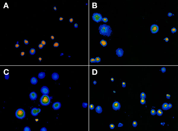
Figure 1. Images of single cell electrophoresis (comet assay) of hRPE cells exposed to microgravity
A: Control cells. B: Cells treated for 24 h with simulated microgravity. These cells had single-stranded DNA breaks as indicated by the appearance of a comet (a cell with a round head and a tail). C: Cells treated for 24 h with simulated microgravity after 48 h recovery. Single-stranded DNA breaks have not been repaired. D: Cells pretreated with 1 μM cysteine, exposed to microgravity for 24 h, and then incubated in media for 48 h recovery. The number of damaged cells was diminished when compared with B,C.