![]() Figure 7 of
Talbot, Mol Vis 2006;
12:65-75.
Figure 7 of
Talbot, Mol Vis 2006;
12:65-75.
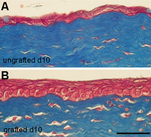
Figure 7. Histological analysis following xenogeneic grafting of human limbal epithelial cells cultured on fibrin gels
Histological corneal staining following xenogeneic grafting, on rabbit, of human limbal epithelial cells cultured on fibrin gels. Masson's Trichrome staining of control rabbits (A; ungrafted) and xenogeneic grafting of cultured human epithelium on rabbit (B). Note that 10 days after grafting, the epithelium regenerated by human epithelial cells is a 5 cell thick layer whereas the ungrafted cornea presents only one or two cell layers containing some goblet cells. The scale bar represents 50 μm.