![]() Figure 6 of
Talbot, Mol Vis 2006;
12:65-75.
Figure 6 of
Talbot, Mol Vis 2006;
12:65-75.
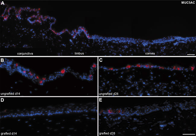
Figure 6. Goblet cell detection in native and grafted rabbit cornea
Goblet cell detection in native rabbit ocular surface in situ and following grafting of rabbit limbal epithelial cells (RLECs) cultured on fibrin gels or controls (ungrafted). Sections were stained with MUC5AC (red), and nuclei were counterstained with Hoechst (blue). Normal rabbit cornea in situ (A) showing the conjunctival, limbal, and corneal areas of the ocular epithelium. Note that the characteristic goblet cell marker, MUC5AC, is increasing with time after surgery in ungrafted corneas from 14 days (B) to 28 days (C). In contrast, the staining was absent or rare in the corneal epithelium 14 days (D) and 28 days (E) after grafting RLECs cultured on the fibrin gel. The scale bars represent 50 μm.