![]() Figure 5 of
Talbot, Mol Vis 2006;
12:65-75.
Figure 5 of
Talbot, Mol Vis 2006;
12:65-75.
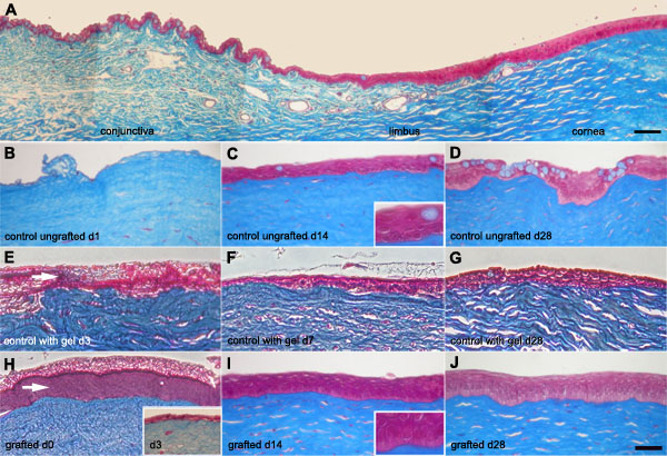
Figure 5. Histological analysis of native and grafted rabbit cornea
Masson's Trichrome staining of native rabbit ocular surface in situ (A) and following grafting of rabbit limbal epithelial cells (RLECs) cultured on fibrin gels (H-J) or controls (ungrafted [B-D] or grafted with fibrin gels without RLECs [E-G]). A: Normal rabbit cornea in situ showing the conjunctival, limbal, and corneal areas of the ocular epithelium. Control corneas (ungrafted [B-D] or grafted with fibrin gels without RLECs [E-G]) or corneas grafted with RLEC cultured on a fibrin gel (H-J) at postoperative day 1 (B), day 3 (E, insert in H), day 7 (F), day 14 (C,I), day 28 (D,G,J) and 2 h after grafting (H). Note that the characteristic staining of goblet cells (a pale blue coloration of round cells) is increasing with time after surgery in control corneas (ungrafted or grafted with fibrin gels without RLECs) as seen at day 1 (B), day 3 (E), day 14 (C), and day 28 (D,G). In contrast, the staining was absent or rare in the corneal epithelium 2 h (H), 14 days (I), and 28 days (J) after grafting RLECs cultured on fibrin gels. Note also that the fibrin gel was present 2 h after grafting (H) but was absent under the epithelium three days post-grafting (insert in H). In contrast, when gels without RLECs were grafted, the fibrin gel was still present three days after grafting (E). The scale bars represent 50 μm.