![]() Figure 4 of
Talbot, Mol Vis 2006;
12:65-75.
Figure 4 of
Talbot, Mol Vis 2006;
12:65-75.
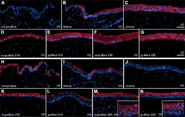
Figure 4. Keratin 3 and 4 analysis of native or grafted rabbit cornea
Keratin 3 (K3) and keratin 4 (K4) staining of native rabbit ocular surfaces in situ and following grafting of rabbit limbal epithelial cells (RLECs) cultured on fibrin gels or controls (ungrafted). A-G: These show immunostaining with K3 (red). H-N: These show immunostaining with K4 (red). A,H: Native conjunctiva. B,I: Native limbus. C,J: Native central cornea. Biopsies harvested from denuded cornea left ungrafted (D,F,K,M) or grafted (E,G,L,N) with autologous limbal epithelium cultured on fibrin for 14 days (D,E,K,L) or 28 days (F,G,M,N). Indirect immunofluorescence staining was performed on frozen sections. Nuclei were counterstained with Hoechst (blue). The scale bars represent 50 μm.