![]() Figure 3 of
Talbot, Mol Vis 2006;
12:65-75.
Figure 3 of
Talbot, Mol Vis 2006;
12:65-75.
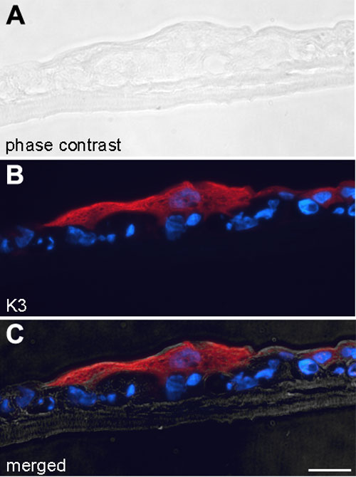
Figure 3. Keratin 3 staining of rabbit limbal epithelial cells cultured on a fibrin gel in vitro
A: Phase contrast of confluent rabbit limbal epithelial cells (RLECs) cultured on fibrin gel. B: Keratin 3 (K3) immunostaining (red) of the same confluent RLECs cultured on fibrin gel. C: A merger of the phase contrast (A) and K3 immunostaining (B). At this step, the RLECs cultured on fibrin gels were ready for grafting. Nuclei were counterstained with Hoechst (blue). The scale bar represents 50 μm.