![]() Figure 2 of
Talbot, Mol Vis 2006;
12:65-75.
Figure 2 of
Talbot, Mol Vis 2006;
12:65-75.
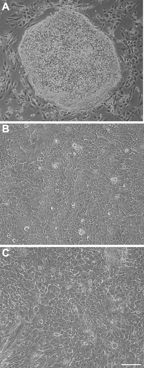
Figure 2. Morphological aspect of rabbit limbal epithelial cells in culture
A: Phase contrast micrographs of (A) a colony of rabbit limbal epithelial cells (RLECs) in primary culture surrounded by fibroblasts from the feeder layer. B: Phase contrast micrographs of confluent cultures (passage 1) of RLECs seeded on plastic. C: Phase contrast micrographs of confluent cultures (passage 1) of RLECs seeded on fibrin gel. The scale bar represents 50 μm.