![]() Figure 1 of
Talbot, Mol Vis 2006;
12:65-75.
Figure 1 of
Talbot, Mol Vis 2006;
12:65-75.
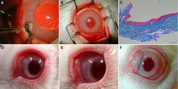
Figure 1. Surgical procedure and grafting
A: A limbectomy was performed over 360° with a round beveled corneal microblade. B: Macroscopic view of the fibrin gel, gently placed over rabbit cornea after complete epithelial removal including limbectomy, and secured with four interrupted sutures in the cardinal positions. C: Microscopic histological analysis of the resected tissue showing that all the epithelial cells were removed after limbectomy. D-F: Macroscopic aspect of the corneal surface after transplantation for ungrafted controls (D; after day 27), grafted with fibrin gel without RLECs (E; day 27) or with RLECs (F; day 23). The scale bar represents 100 μm.