![]() Figure 2 of
Konopatskaya, Mol Vis 2006;
12:626-632.
Figure 2 of
Konopatskaya, Mol Vis 2006;
12:626-632.
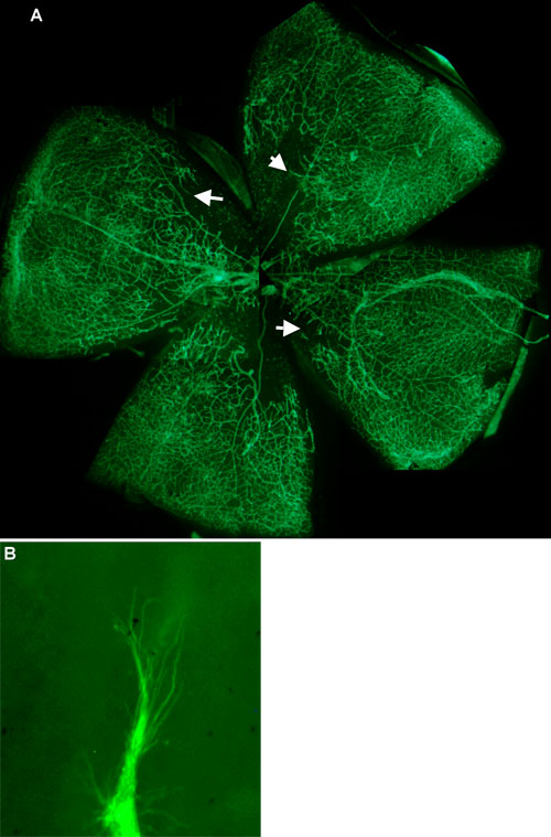
Figure 2. FITC-Isolectin staining of hypoxic retina after injection with 1 ng VEGF165b
A: Although significant hypoxia is still apparent in the retinal whole mount (approximately 4.6 mm across), vascular abnormalities are rare, and neovascularization appears to be proceeding into the area of ischemia along the plane of the retina (arrows; 4x objective). B: Sprouts, 20-40 μm long, are still seen at the edge of the ischemic area, but in the plane of the retina (100x objective).