![]() Figure 1 of
Konopatskaya, Mol Vis 2006;
12:626-632.
Figure 1 of
Konopatskaya, Mol Vis 2006;
12:626-632.
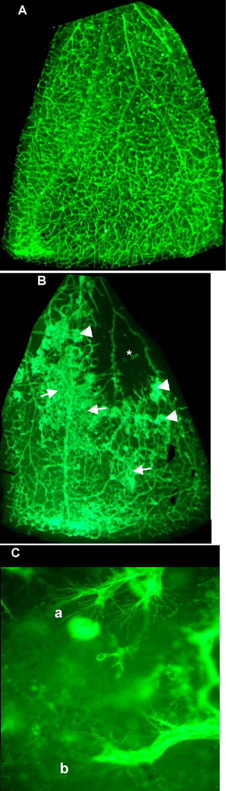
Figure 1. FITC-Isolectin staining of retina in normoxic and relative hypoxic mice
A: Each quadrant of the normoxic retinal whole mounts is approximately 2.2 mm across. B: Relative hypoxia was induced by incubation in 95% oxygen during vascular development followed by return to normoxia at day 12. Ischemic area is indicated by an asterisk (*). Neovascular areas are characterized by vascular abnormalities (arrowhead), indistinct vascular outlines, and "fuzziness" (arrows). C: High power view of neovascular area showing sprouting endothelial cells with filopodia extending 20-30 μm both within (a) and outside (b) the plane of focus. A,B: 4x objective. C: 63x objective.