![]() Figure 1 of
Prow, Mol Vis 2006;
12:616-625.
Figure 1 of
Prow, Mol Vis 2006;
12:616-625.
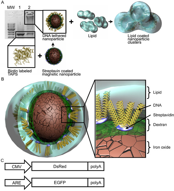
Figure 1. Construction and anatomy of magnetic nanoparticles
A: Conjugation of biotin-labeled transcriptionally active PCR products (TAP) DNA to streptavidin-coated magnetic nanoparticles (MNP). A 0.8% agarose gel stained with ethidium bromide was used to visualize DNA and DNA tethered nanoparticles. The leftmost lane are molecular weight markers, from 1 to 10 kb (MW). Lane 1 contains only 5' biotin-tagged TAP DNA (Black rectangular outline). Lane 2 is a solution containing DNA from Lane 1 combined with streptavidin-coated magnetic nanoparticles. The black rectangular outline in Lane 2 highlights 5' TAP tethered magnetic nanoparticles. A: Schematic of the construction of the MNP. B: The layered anatomy of a lipid coated DNA tethered nanoparticle. C: Schematics of the two DNA constructs used to assess transfection and ARE activity.