![]() Figure 6 of
Prow, Mol Vis 2006;
12:606-615.
Figure 6 of
Prow, Mol Vis 2006;
12:606-615.
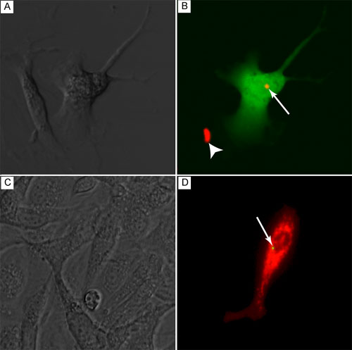
Figure 6. Lipid-coated nanocrystal transfected human retinal epithelium cells
Cells were cultured with lipid-coated nanocrystals tethered to either EGFP (green in B) or DsRed (red in D) for 48 h or 10 days, respectively. Confocal (A,B) and fluorescence (C,D) microscopy were used to simultaneously visualize nanocrystals and tethered fluorescent gene expression. The nanocrystals are marked by white arrows and nanocrystal aggregate is marked with a white arrowhead. A: DIC microscopy. C: Phase contrast microscopy.