![]() Figure 2 of
Prow, Mol Vis 2006;
12:606-615.
Figure 2 of
Prow, Mol Vis 2006;
12:606-615.
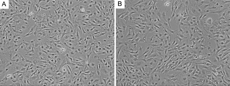
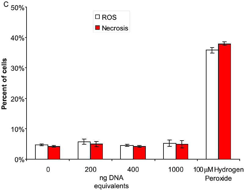
Figure 2. Adult dog retinal epithelium cells cytotoxicity from uncoated magnetic nanoparticles
The images are representative phase contrast photomicrographs of ADREC treated with 0 ng (A) or 400 ng (B) DNA tethered MNP without lipid-coating for 24 h. C: The effects of uncoated magnetic nanoparticles (0, 200, 400, 1000 ng DNA equivalents) on reactive oxygen species (ROS; white bars) and necrosis (red bars) after 48 h incubation. 100 μM tert-butyl hydrogen peroxide was used as a positive control. The error bars represent standard deviation. One potential cause for concern when using iron derived nanoparticles is iron induced oxidative stress, while another concern may be membrane integrity. Therefore, an oxidative stress sensitive fluorescent dye was used to confirm the absence of nanoparticle induced oxidative stress. Likewise, propidium iodide was used to confirm the absence of ruptured membrane. Flow cytometric analysis of these cells confirmed that there was no increase in oxidative stress or ruptured membranes (Necrosis).