![]() Figure 4 of
Szabo, Mol Vis 2006;
12:597-605.
Figure 4 of
Szabo, Mol Vis 2006;
12:597-605.
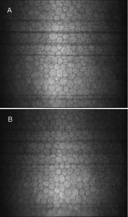
Figure 4.
In vivo confocal microscopic images of the endothelium of untreated healthy cornea (A) and the endothelium of PRK-treated cornea with clinically significant subepithelial haze (B), showing no difference in endothelial morphology and cell density.