![]() Figure 3 of
Szabo, Mol Vis 2006;
12:597-605.
Figure 3 of
Szabo, Mol Vis 2006;
12:597-605.
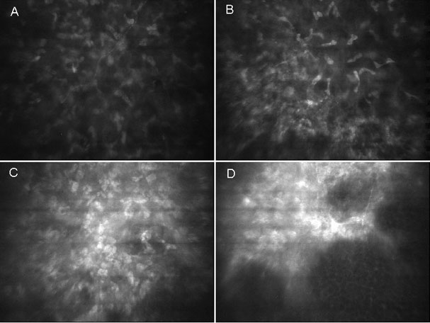
Figure 3.
In vivo confocal microscopic images of untreated healthy cornea (A), PRK-treated cornea with normal wound healing (B), PRK-treated cornea with mild haze and activated keratocytes (C), and PRK-treated cornea with clinically significant subepithelial haze (D). Images were obtained within the anterior 50 μm of the stroma.