![]() Figure 4 of
Sreelakshmi, Mol Vis 2006;
12:581-587.
Figure 4 of
Sreelakshmi, Mol Vis 2006;
12:581-587.
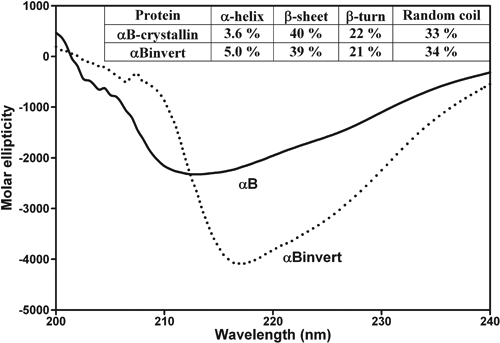
Figure 4.
Far-UV CD spectra of wild-type αB- and αBinvert-crystallin mutant. Spectra were recorded at a protein concentration of 0.2 mg/ml using a 0.05 cm path length cell. Contents of secondary structural elements were determined using the CDPro suite of programs [23]. When compared to wild-type αB-, αBinvert-crystallin mutant exhibited a 1.4% increase in α-helical content at the expense of 1% reduction each in β-sheet and β-turn contents.
