![]() Figure 9 of
Lutty, Mol Vis 2006;
12:532-580.
Figure 9 of
Lutty, Mol Vis 2006;
12:532-580.
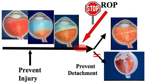
Figure 9. Rationale for the STOP-ROP study
Artist's concept of the sequential appearance of the growing preterm human retina as it develops ROP. From left to right, the normal immature retina, initial injury of the developing vessels, neovascularization with plus disease, involution (top) or retinal detachment (lower). The red arrow and circle indicates the hypothesized time/site of action of oxygen in the STOP-ROP study.