![]() Figure 8 of
Lutty, Mol Vis 2006;
12:532-580.
Figure 8 of
Lutty, Mol Vis 2006;
12:532-580.
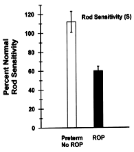
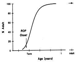
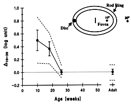
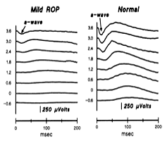
Figure 8. Visual function and ROP
A: The growth curve summarizes the developmental increase in human rhodopsin. ROP has its onset just as the curve begins to increase rapidly. B: The mean and prediction limits for D 10-30 in normal infants; the adult value of zero is reached by age 6 months. C: Sample ERG records from 10-week-old infants. The vertical scale is in log scotopic troland seconds. The downward deflections in the records are the a-waves (arrow). The difference between records from the infant with mild ROP and the normal subject are conspicuous. D: The a-waves are displayed on an expanded time scale. The dashed lines represent the Lamb & Pugh model fitted to the ERG a-waves. S (rod cell sensitivity) is lower in ROP subjects. E: The mean values of S for "No ROP" and "ROP" subjects are shown as percent of normal for age. The means differ significantly (re-plotted from Fulton et al, 2001).
A: Republished with permission of The Association for Research in Vision & Ophthalmology from Fulton AB, Dodge J, Hansen RM, Williams TP. The rhodopsin content of human eyes. Invest Ophthalmol Vis Sci 1999; 40:1878-83. B: Republished with permission of The Association for Research in Vision & Ophthalmology from Hansen RM, Fulton AB. The course of maturation of rod-mediated visual thresholds in infants. Invest Ophthalmol Vis Sci 1999; 40:1883-6. C,D: Republished with permission of the American Medical Association from Fulton AB, Hansen RM, Petersen RA, Vanderveen DK. The rod photoreceptors in retinopathy of prematurity: an electroretinographic study. Arch Ophthalmol 2001; 119:499-505. Copyright ©2001 American Medical Association. All rights reserved.
A:
B:
C:
D:

E: