![]() Figure 2 of
Lutty, Mol Vis 2006;
12:532-580.
Figure 2 of
Lutty, Mol Vis 2006;
12:532-580.
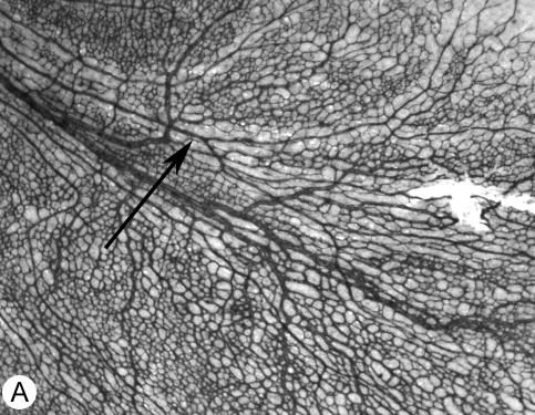
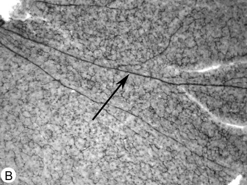
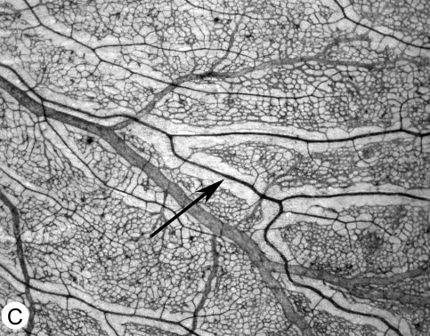
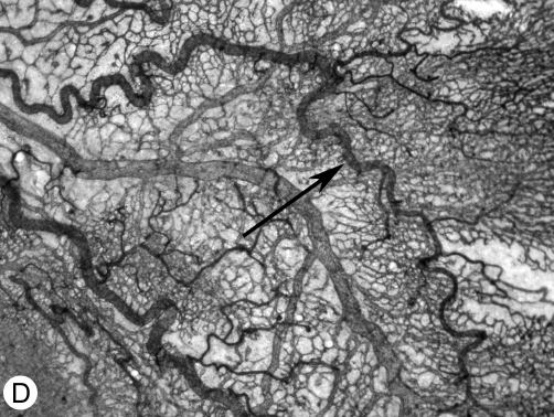
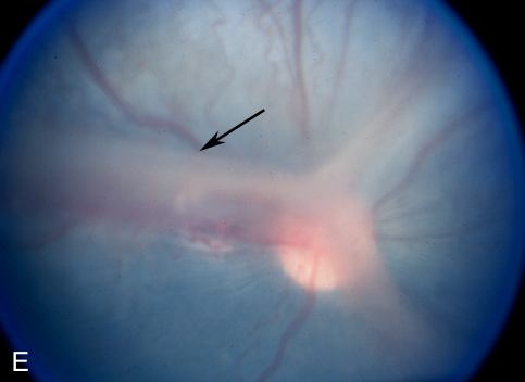
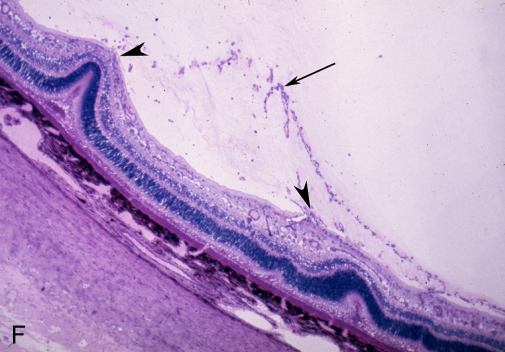
Figure 2. Canine model of oxygen-induced retinopathy
A: ADPase whole mount showing the temporal arcade of a 5-day-old normal air control dog (the arrow indicates an artery). Note the density and chicken wire mesh appearance of the developing capillaries. B: ADPase whole mount showing the temporal arcade of a 5-day-old dog sacrificed after 4 days of 100% oxygen exposure (the arrow indicates an artery). Note the obliteration of the capillaries and the extreme constriction of the major vessels. C: ADPase whole mount showing the temporal arcade of a 21-day-old normal air control dog (the arrow indicates an artery). Capillary-free zones have formed along the arteries and remodelling of the capillary bed has occurred. The secondary capillary bed has also formed. D: ADPase whole mount showing the temporal arcade of a 21-day-old oxygen-treated dog after return to room air for 16 days. The arteries are extremely tortuous (arrow) and an overgrowth of the capillaries is apparent. E: Fundus photograph of a 45-day-old oxygen-treated dog. Preretinal membranes extend along the major arcades and over the optic nerve head (arrow). F: Histological section from the animal shown in Panel E, shows tractional retinal folds (arrowheads) associated with the preretinal membrane (arrow).