![]() Figure 16 of
Lutty, Mol Vis 2006;
12:532-580.
Figure 16 of
Lutty, Mol Vis 2006;
12:532-580.
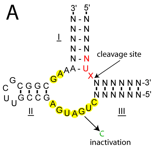
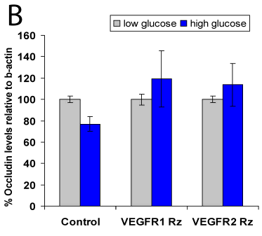
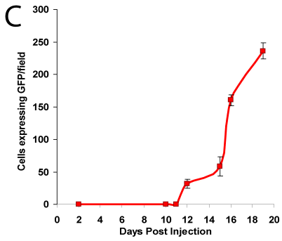
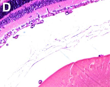
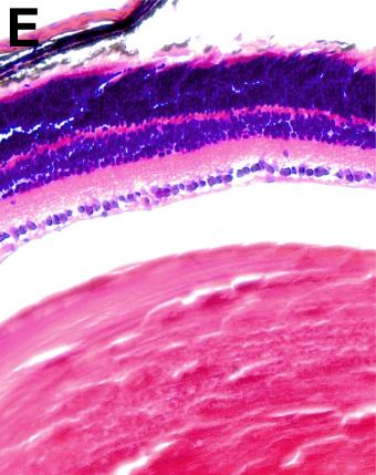
Figure 16. Targeted ribozyme expression vectors in an OIR mouse model
A: Structure of a generic hammerhead ribozyme annealed to its target. Cleavage occurs just downstream of the target NUX site where N is any base and X is not a G. Bases highlighted in yellow show the catalytic core. B: HRECs transfected with plasmids expressing ribozymes targeting vascular endothelial growth factor receptors 1 and 2 (VEGFR-1, VEGFR-2) prevent reduction in occludin levels in high glucose (25 mM). C: Intraocular injection of 1 μg of plasmid expressing green fluorescent protein (GFP) into the oxygen-induced retinopathy (OIR) mouse model on postnatal day 1 results in increasing GFP expression on postnatal day 12 through 19. D: Cross section of control retina from mouse from the OIR model showing preretinal neovascularization. E: Cross section of retina from mouse from the OIR model after intraocular injection on postnatal day 1 with a plasmid expressing IGF-1R ribozyme. Note lack of preretinal neovascularization.