![]() Figure 10 of
Lutty, Mol Vis 2006;
12:532-580.
Figure 10 of
Lutty, Mol Vis 2006;
12:532-580.
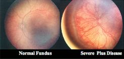
Figure 10. Fundus photography of plus disease in ROP
Photographs of the posterior pole of human infants demonstrating the normal vascular pattern (left), and venous dilation and arteriolar tortuosity of "plus disease" (right). RetCam photos courtesy of Massie Labs (now Clarity Medical Systems, Inc.), Pleasanton, CA.