![]() Figure 3 of
Yang, Mol Vis 2006;
12:511-517.
Figure 3 of
Yang, Mol Vis 2006;
12:511-517.
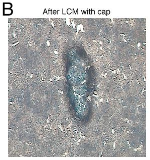
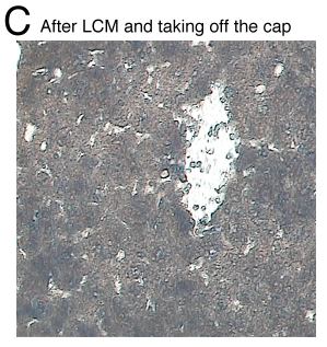
Figure 3. Laser capture microdissection (LCM) of hepatic micrometastatic melanoma
A: Hepatic micrometastasis in a mouse hepatic frozen section prior to LCM. B: Laser spots outline the micrometastasis. The micromestastasis cells are inside the border. The normal hepatocytes outside the border are not disturbed. C: After the laser spot outline the micrometastasis, the tissue is removed by the cap. Approximately 90% of the cells in the micrometastasis are captured.