![]() Figure 3 of
John-Aryankalayil, Mol Vis 2006;
12:55-64.
Figure 3 of
John-Aryankalayil, Mol Vis 2006;
12:55-64.
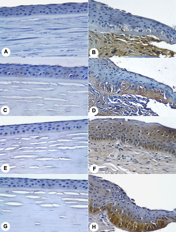
Figure 3. Immunohistochemical analysis of proteins in pterygium and normal cornea
Immunohistochemical staining for fibronectin (A,B), lipocalin 2 (C,D), MIP-4 (E,F), and L-PGDS (G,H) was increased in pterygium (right panels) when compared to normal cornea (left panels). B: Abundant fibronectin staining occurs along the entire length of the pterygium stroma and in some stromal fibroblasts. C: Lipocalin 2 is faintly detected in normal corneal basal epithelial cells. D: Lipocalin 2 immunoreactivity is localized to pterygial basal epithelial cells, basement membrane, and some extracellular components of the stroma. F: MIP-4 is present in both the stroma and the epithelial cell layers. H: Intense staining of L-PGDS was observed in the basal epithelial cell layer. A thinly stained line at the pterygial and corneal cell surface may either be a specific reaction by terminally differentiated epithelial cells or a possible immunological edge effect. Protein localization in the specified areas was the same in the wider field seen under lower magnification.