![]() Figure 4 of
Shimmura, Mol Vis 2006;
12:478-484.
Figure 4 of
Shimmura, Mol Vis 2006;
12:478-484.
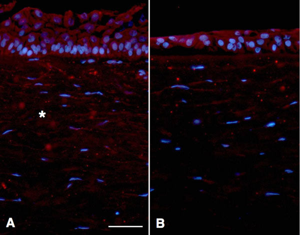
Figure 4.
Immunohistology of a corneolimbal segment using an anti-SPARC monoclonal antibody. The subepithelial tissue in the limbus (A) showed SPARC distributed in the interstitial space (asterisk). SPARC-associated signals were much lower in the central cornea (B). The scale bar represents 50 μm.