![]() Figure 1 of
Shimmura, Mol Vis 2006;
12:478-484.
Figure 1 of
Shimmura, Mol Vis 2006;
12:478-484.
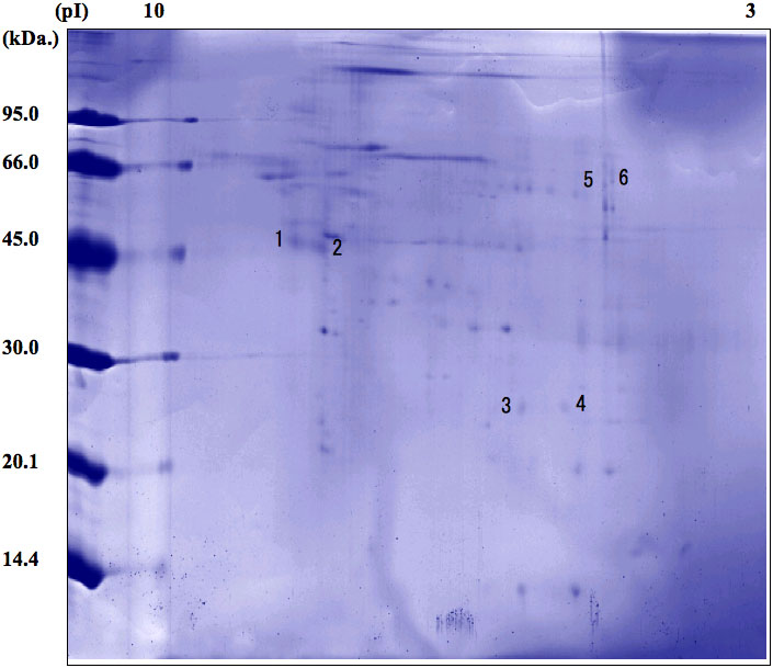
Figure 1.
2-D PAGE of proteins concentrated from the supernatant of limbal fibroblasts. Spots were visualized with Coomassie brilliant blue. Six spots (numbered) were found to be distinctively expressed by limbal fibroblasts and were identified by ion-trap mass spectrometry. The name and GenBank accession number for each protein are listed in Table 1.