![]() Figure 5 of
Davies, Mol Vis 2006;
12:467-477.
Figure 5 of
Davies, Mol Vis 2006;
12:467-477.
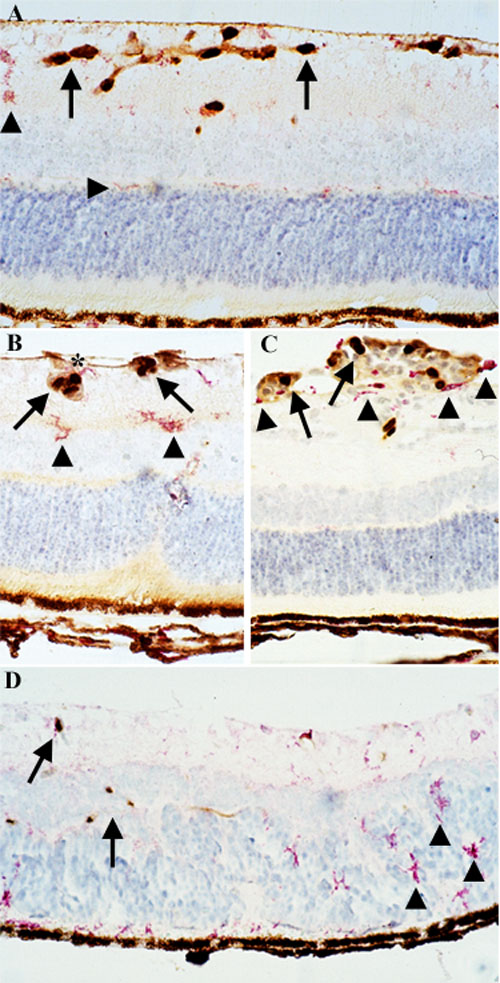
Figure 5. Retinal cell proliferation in oxygen-induced retinopathy
P14O2 retinas (A) showed an increase in BrdU+ nuclei (brown, arrows) following oxygen-induced injury, with F4/80+ cells (red, arrowheads) localized to the outer plexiform layer, inner plexiform layer, and ganglion cell layer (GCL). P17O2 retinal cross sections stained for F4/80 and BrdU shows rare co-localization (asterisk) of microglia and macrophages (MG/MAC; arrowheads) and proliferating nuclei (arrows; B,C). P21O2 retinal cross sections (D) showed that MG/MAC located in deeper nuclear layers of the retina (arrowheads) did not correspond to those cells that are actively proliferating (BrdU+, arrows).