![]() Figure 4 of
Davies, Mol Vis 2006;
12:467-477.
Figure 4 of
Davies, Mol Vis 2006;
12:467-477.
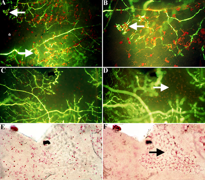
Figure 4. Microglia and macrophages localization in P21O2 retinas
In the P21O2 central retina, microglia and macrophages (red) continue to be primarily localized to vascular and avascular transition regions (A, asterisk) and neovascular tufts (A, arrow). Higher magnification (A, arrow) again reveals the intimate relationship between microglia and macrophages (MG/MAC) and neovascular tufts (B, arrow). While in the peripheral retina, MG/MAC are also localized to the superficial vascular network of the retina (C,E). However, in the deep vascular network MG/MAC are localized in areas corresponding to the avascular region (D,F, arrows). Magnification 100x (A), 200x (B-F).