![]() Figure 3 of
Davies, Mol Vis 2006;
12:467-477.
Figure 3 of
Davies, Mol Vis 2006;
12:467-477.
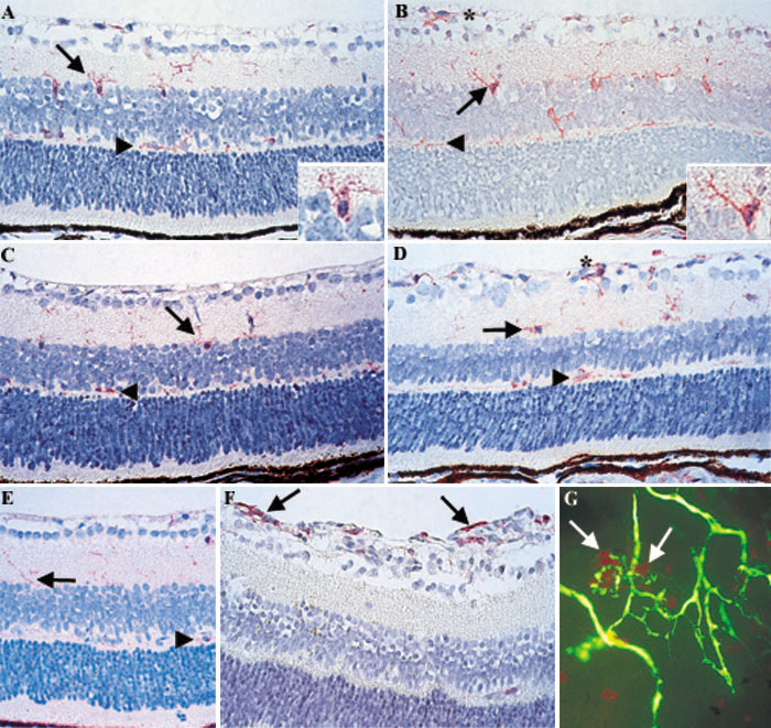
Figure 3. Localization of microglia and macrophages in oxygen-induced retinopathy
A: Immunostaining for F4/80 in P12 room air retinas revealed F4/80+ cells (red) along the inner plexiform layer (IPL; arrow) and outer plexiform layer (OPL; arrowhead). F4/80+ cells located in the IPL (arrow) are seen around pyknotic nuclei (insert). B: P12O2 retinas showed a similar staining pattern with cells along both the IPL (arrow) and OPL (arrowhead), with additional cells along the nerve fiber layer (NFL, asterisk). As with P12 room air- control retinas, F4/80+ staining is localized to pyknotic cells (insert) located in the IPL (arrow). C: P14 control retinas have comparable staining to P12 room air- retinas (IPL, arrow; OPL, arrowhead). D: Following oxygen-induced injury, P14O2 retinas demonstrated increased F4/80+ cells along the NFL (asterisk) in addition to those cells found in the IPL (arrow) and OPL (arrowhead). E: P17 room air controls have slightly less activated F4/80+ cells still localized to the IPL (arrow) and OPL (arrowhead). F: In contrast, staining of P17O2 retinas revealed almost no staining along either plexiform layer of the retina, and numerous F4/80+ cells located within the neovascular tufts (arrows). G: Fluorescein angiography combined with F4/80 immunostaining demonstrates the intimate relationship between microglia and macrophages (red) and vessels (green) in the neovascular tufts (arrows). Magnification 400x.