![]() Figure 2 of
Davies, Mol Vis 2006;
12:467-477.
Figure 2 of
Davies, Mol Vis 2006;
12:467-477.
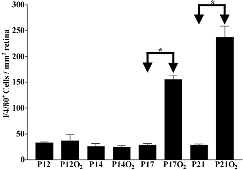
Figure 2. Quantification of retinal microglia and macrophages in oxygen-induced retinopathy
F4/80+ cell numbers increased on P17O2 and P21O2 compared to age-matched room air controls (O2 refers to hyperoxia exposure from P7 to P12 only). Cell numbers remained constant at all other times and conditions examined. Asterisk indicates p<0.001 by one-way ANOVA followed by the Tukey's post-test.