![]() Figure 1 of
Davies, Mol Vis 2006;
12:467-477.
Figure 1 of
Davies, Mol Vis 2006;
12:467-477.
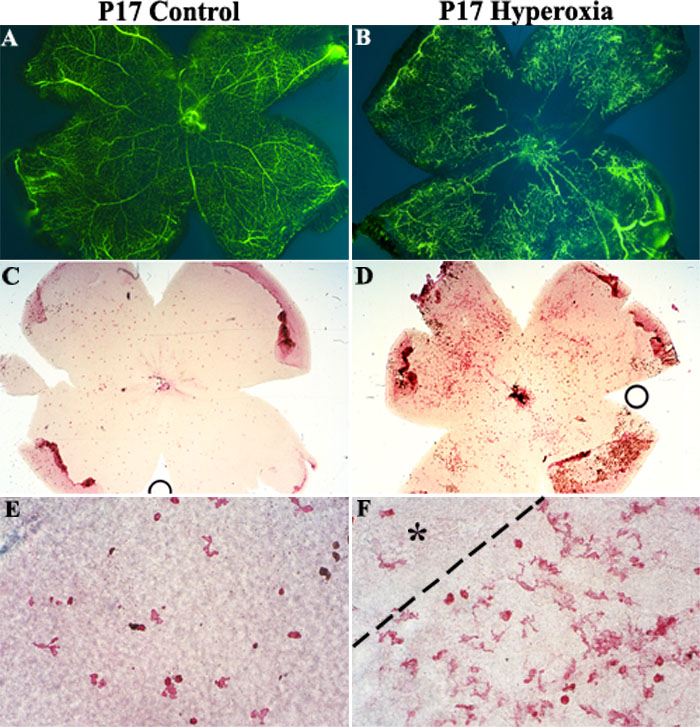
Figure 1. Immunolabeled F4/80 cells in retinal flat mounts
Fluorescence angiography showed retinal vasculature in B6 mice on P17 room air (A) compared to P17 Hyperoxia mice (B). Immunohistochemical localization of F4/80+ cells in P17 control retinas (C) compared to P17 Hyperoxia retinas (D). Higher magnification of F4/80+ cells in P17 control (E) and P17 Hyperoxia (F) retinas revealed not only an increased number of microglia/macrophages but also more activated microglia with multiple processes. Dashed line (F) denotes the transition region between avascular (asterisk) and vascular retina where neovascularization is known to occur. Magnification 25x (A-D); 400x (E-F).