![]() Figure 4 of
El Kochairi, Mol Vis 2006;
12:461-466.
Figure 4 of
El Kochairi, Mol Vis 2006;
12:461-466.
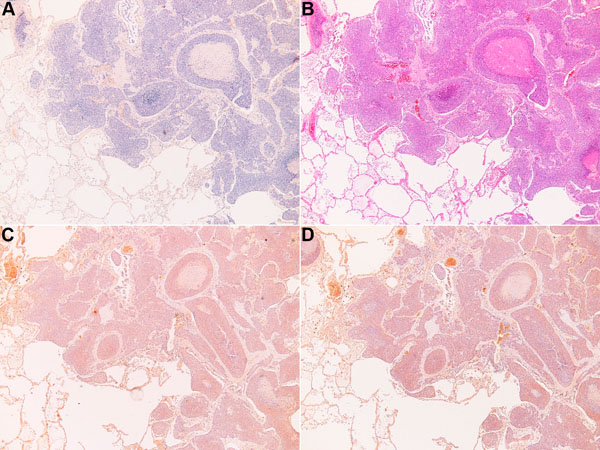
Figure 4. Absence of keratoepithelin deposits in the squamous cell carcinoma
A: Congo red staining showed no amyloid deposits. B: Hematoxylin and eosin (H&E) staining of squamous cell carcinoma tissue revealed no deposit. C: Control preimmune serum treatment on squamous cell carcinoma. D: Immunostaining with KE2 does not reveal any deposit in the tumoral pulmonary tissue.The brownish spots correspond to blood red cells.