![]() Figure 3 of
El Kochairi, Mol Vis 2006;
12:461-466.
Figure 3 of
El Kochairi, Mol Vis 2006;
12:461-466.
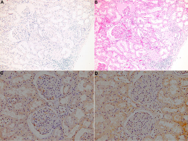
Figure 3. Absence of keratoepithelin deposits in the kidney
A: Congo red staining showed no amyloid deposits. B: Hematoxylin and eosin (H&E) staining of kidney tissue revealed no deposit. C: Control preimmune serum treatment on kidney tissue. D: Immunostaining with KE2 is uniformally positive because the kidney normally expresses keratoepithelin, but it is negative for pathological deposits of this protein that are characteristic of TGFBI/BIGH3-related corneal dystrophies. The brownish spots correspond to blood red cells.