![]() Figure 1 of
El Kochairi, Mol Vis 2006;
12:461-466.
Figure 1 of
El Kochairi, Mol Vis 2006;
12:461-466.
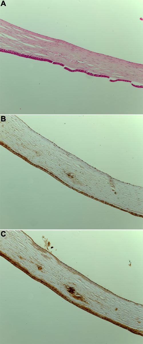
Figure 1. Keratoepithelin deposit in the patient's cornea (LCDI)
A: Hematoxylin and eosin (H&E) staining of the cornea showed deposits that are disturbing the normal architecture of the corneal stroma. B: Control preimmune serum treatment on the cornea. C: Immunostaining with keratoepithelin antibody (KE2) revealed pathological intrastroma deposits of keratoepithelin. Corneal epithelium is uniformally stained with KE2 because it normally expresses keratoepithelin. In the images, KE refers to keratoepithelin and KE2 refers to antibody raised against KE.