![]() Figure 1 of
Karring, Mol Vis 2006;
12:451-460.
Figure 1 of
Karring, Mol Vis 2006;
12:451-460.
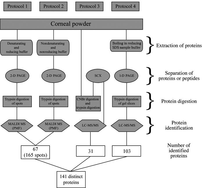
Figure 1.
Schematic diagram showing the four different protocols used for the extraction, separation, and identification of proteins from the human cornea. The right side of the figure characterizes each step of the procedures.