![]() Figure 3 of
Yao, Mol Vis 2006;
12:445-450.
Figure 3 of
Yao, Mol Vis 2006;
12:445-450.
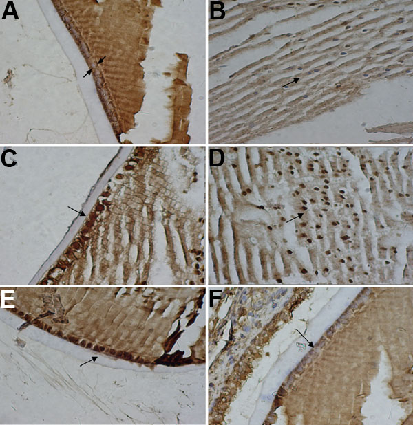
Figure 3. Micrographs showing the distribution of Hsp70 immunoreactivity in rat lens epithelium
Immunostaining of normal eyes revealed Hsp70 was negative in lens epithelial cells (A) and the fiber cells of the lens (B) and is highlighted by arrows. Hsp70 reactivity in contusion eyes indicated it was localized in the nucleus or cytoplasm around the nucleus, and exhibited increased immunostaining in the lens epithelieal cells (C) and the fiber cells of the lens (D) and is highlighted by arrows. Hsp70 is expressed strongly in lens epithelial cells in the heat shock eye (E), and faintly in lens epithelial cells in the quercetin eye (F). Magnification was 400x.