![]() Figure 1 of
Yao, Mol Vis 2006;
12:445-450.
Figure 1 of
Yao, Mol Vis 2006;
12:445-450.
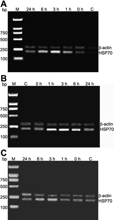
Figure 1. Expression of Hsp70 mRNA in rat lens epithelial cells
In the contusion group (A), Hsp70 is expressed normally but increased progressively by 1 h after eye trauma, peaked at 3 h, and decreased progressively by 24 h. In the heat shock group (B), Hsp70 was expressed higher after heat shock, increased progressively by 1 h, peaked at 3 h, and decreasing at 24 h but still remained higher than the control subgroup. In the quercetin group (C), Hsp70 also increased at 1 h and peaked at 3 h, then decreased progressively until 24 h, but the densitometry value of each subgroup was lower than the control group. D: Comparison of three groups shows the heat shock group was the highest and the quercetin group was the lowest. M indicates the molecular weight marker. The lanes are identified as control (C) or by the duration until enucleation for the subgroup within each group.