![]() Figure 3 of
Layton, Mol Vis 2006;
12:43-54.
Figure 3 of
Layton, Mol Vis 2006;
12:43-54.
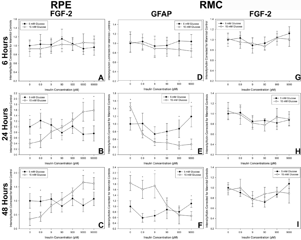
Figure 3. The effect of insulin on retinal pigment epithelium (RPE) and Müller cell protein expression
The effect of insulin on RPE and Müller cell protein expression was measured by immunoblotting, in conditions of high (15 mM) and low (5 mM) glucose. Results are expressed as a proportion of the expression of the substance in 5 mM glucose in the absence of insulin. A,B,C: The effect of insulin on fibroblast growth factor-2 (FGF-2) production in RPE culture. After 6 h, insulin and glucose levels have no effect on RPE FGF-2 expression. However, after 24 h, FGF-2 production is significantly decreased (p<0.01) in conditions of low insulin but only in the 15 mM glucose environment. Conversely, high insulin levels increased FGF-2 production in the RPE significantly in the 15 mM glucose environment relative to the 5 mM environment (p<0.01). This effect is maintained in other cultures analyzed 48 h after treatment. In a 5 mM environment, insulin levels in this range do not alter FGF-2 expression in cultured RPE cells at any of the times tested. D,E,F: Similar experiments in cultured Müller cells are shown. GFAP expression in Müller cells is unaffected by insulin or glucose treatments after 6 h, however, after 24 h of treatment, very low levels of insulin are noted to increase GFAP expression significantly in an environment of 15 mM glucose (p<0.01). GFAP expression is decreased with increasing insulin concentrations in the 15 mM environment and is significantly reduced relative to the 5 mM environment after treatment with 900 pM and 9 nM of insulin (p<0.05). After 48 h of treatment in other cultures, low insulin levels increased GFAP levels to a significantly greater degree (p<0.05 at the lowest insulin levels), and the increase was significant relative to the 5 mM environment across a greater range of concentrations. After 48 h in the 15 mM glucose environment, 9 nM insulin was still effective in reducing GFAP expression both relative to the insulin free conditions (p<0.01) and to all treatments in the 5 mM environment. In the environment of 5 mM glucose, GFAP expression was not significantly altered with insulin treatment across the range of concentrations and treatment durations tested here. G,H,I: FGF-2 production in cultured Müller cells was not significantly altered by insulin in either glucose environment across the range of concentrations and treatment durations investigated here. Asterisk indicates p<0.05 by Student's unpaired t-tests comparing protein expression in the 5 and 15 mM glucose environments with five independent samples for each treatment condition and at each time.