![]() Figure 1 of
Layton, Mol Vis 2006;
12:43-54.
Figure 1 of
Layton, Mol Vis 2006;
12:43-54.
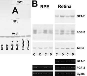
Figure 1. The effects of diabetes on FGF-2 and GFAP
The effect of 15 weeks of diabetes on retinal and retinal pigment epithelium fibroblast growth factor-2 and retinal GFAP expression was measured by immunoblotting. A: Immunoblotting for neurofilament light (NFL, an inner retinal marker), von Willebrand Factor (vWF; monomer, an endothelial marker), and actin (a housekeeping structural protein), showing the method used to produce RPE samples for analysis yielded relatively little contamination from the retina or choroid. B: Representative immunoblots showing upregulation of FGF-2 and GFAP protein and mRNA in the diabetic retina but downregulation of FGF-2 protein and mRNA in the diabetic RPE. The control (C) and diabetic (D) lanes are identified. Cyclophilin is abbreviated "Cyclo". C: FGF-2 protein and mRNA was reduced by 25±6% (p<0.01) and 35±4% (p<0.01) in the diabetic RPE. In contrast, retinal FGF-2 protein and mRNA were increased by 26±7% (p<0.01) and 47±8% (p<0.01). Similarly, GFAP expression was also increased in the diabetic retinas. Asterisk (*) indicates p<0.01 after accounting for intra- and interclass variability between eyes and within animals by repeated measures analysis of variance; 18 eyes were studied in each group. D: Retinal FGF-2 protein levels were not related to the FGF-2 expression in the corresponding eye's RPE (p<0.87). E: Retinal GFAP levels were correlated to the FGF-2 expression in the corresponding eye's RPE (r2=0.31, p<0.01). Dotted lines indicate 95% prediction intervals of the regression. Results are expressed proportionally in relation to 5 mM glucose in the absence of insulin.