![]() Figure 4 of
Chung, Mol Vis 2006;
12:415-421.
Figure 4 of
Chung, Mol Vis 2006;
12:415-421.
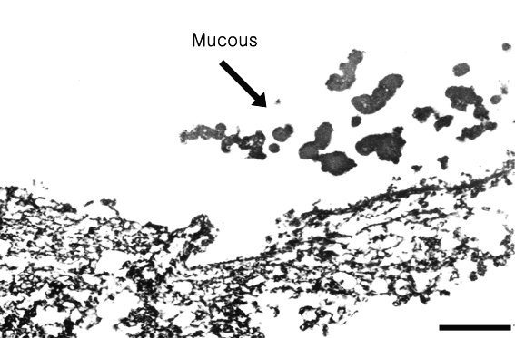
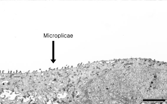
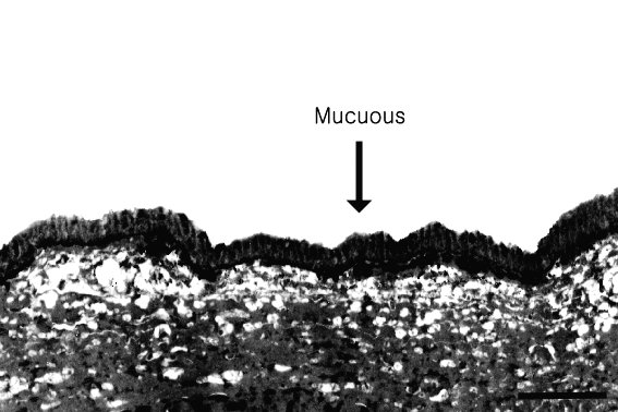
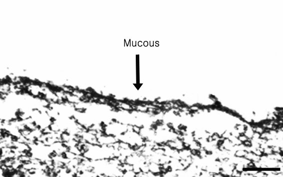
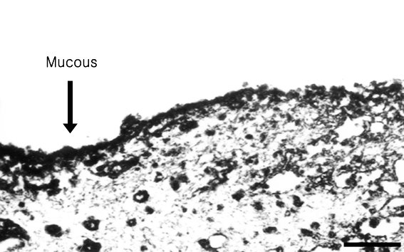
Figure 4. Transmission electron microscopy
Transmission electron micrographs of human corneas treated with 0.01% BAC solution. A: The negative control cornea (no BAC) showed no mucous layer. B: The positive control cornea fixed with CPC showed a well defined mucous layer. C: Cornea treated with BAC solution for 5 min showed a thin trace of mucous layer on the surface. D: Corneas treated with BAC solution for 15 min showed a thinned mucous layer. E: Corneas treated with BAC solution for 60 min had a near total loss of the mucous layer. The mucous present was mostly seen clumped near the epthelium, but no longer attached to it. Scale bars represent 1 μm.
A:
B:
C:
D:
E: