![]() Figure 5 of
Cryan, Mol Vis 2006;
12:405-414.
Figure 5 of
Cryan, Mol Vis 2006;
12:405-414.
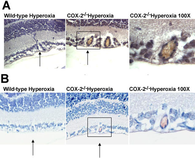
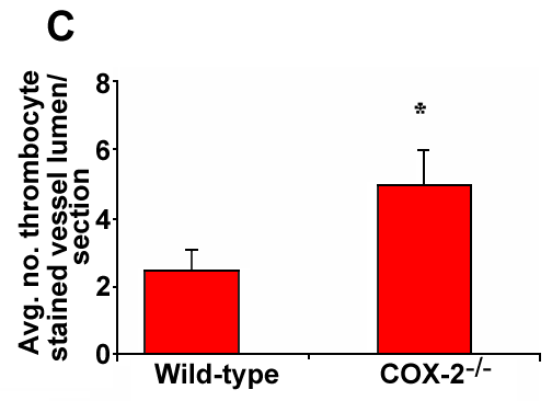
Figure 5. Immunohistochemical detection of thrombosis in retinal blood vessels of hyperoxia-treated mice
Mice were exposed to 75% oxygen from postnatal days 7 to 12 and subsequently to normoxic conditions for a further five to six days (as described in Materials and Methods). Both fibrin and thrombocytes were detected immunohistochemically in the retinal vessels of these mice as described in the Materials and Methods section. A: Fibrin staining of hyperoxia-treated wild-type and COX-2-/- retinas. B: Thrombocyte staining of hyperoxia-treated wild-type and COX-2-/- retinas. Magnification was 40x. Arrows indicate the presence of retinal blood vessel lumen. C: Quantitative analysis of thrombocyte-stained lumen in hyperoxia-treated wild-type and COX-2-/- retinas. Results are means; error bars represent SEM. There were at least six animals per group. Asterisks indicates comparison was statistically significant (p<0.05).