![]() Figure 4 of
Cryan, Mol Vis 2006;
12:405-414.
Figure 4 of
Cryan, Mol Vis 2006;
12:405-414.
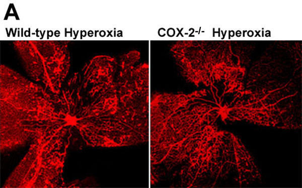
Figure 4. Isolectin B4 stained retinal vasculature in wild-type and COX-2-/- mice at postnatal day 18, following oxygen-induced retinopathy
A: Isolectin B4 stained retinas captured using confocal scanning laser microscopy (magnification 4x). B: Quantification of the percentage area of retina where neovascular tufts were present in lectin-stained retinas from P18 hyperoxia-treated wild-type and COX-2-/- mice using Angiostat. C: Quantification of the percentage area of capillary-free retina in lectin-stained retinas from P18 hyperoxia-treated wild-type and COX-2-/- mice using Angiostat. Results are means; error bars represent SEM. There were at least 6 animals per group. The comparisons marked "NS" were not statistically significant.