![]() Figure 3 of
Cryan, Mol Vis 2006;
12:405-414.
Figure 3 of
Cryan, Mol Vis 2006;
12:405-414.
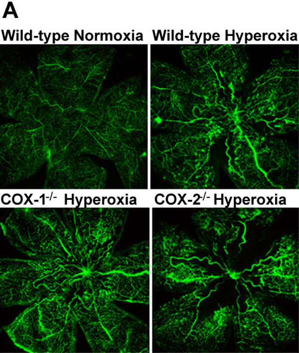
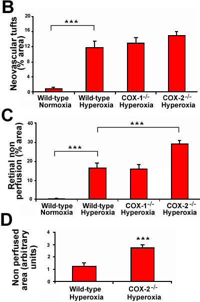
Figure 3. Fluorescein perfused retinal vasculature following oxygen-induced retinopathy in COX knockout mice
Mice were exposed to 75% oxygen from postnatal days 7 to 12 and subsequently to normoxic conditions for a further six days (as described in Materials and Methods). A: Deeply anesthetized mice were perfused with fluorescein-dextran, and their retinas flatmounted as described in the Materials and Methods section (magnification 4x). B: Quantification of the percentage area of the retina where neovascular tufts were present, in each fluorescein-perfused group at postnatal day 18 (P18) using the program, Angiostat. C: Quantification of the percentage area of retinal nonperfusion present in fluorescein-perfused retinal flatmounts at P18 using Angiostat. D: Quantification of the area of nonperfused retina, in the central area of interest, in wild-type and COX-2-/- hyperoxia-treated fluorescein-perfused P18 mice using Image Pro Plus. Results are means; error bars represent SEM. There were at least six animals per group. Asterisks indicate comparisons were statistically significant (p<0.001).