![]() Figure 2 of
Cryan, Mol Vis 2006;
12:405-414.
Figure 2 of
Cryan, Mol Vis 2006;
12:405-414.
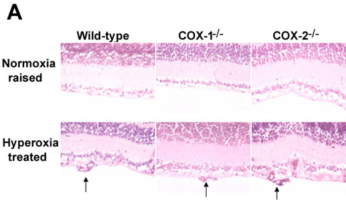
Figure 2. The effect of disrupting the COX-1 or COX-2 gene on retinal morphology and intravitreal endothelial nuclei in a mouse model of oxygen-induced retinopathy
Mice were exposed to 75% oxygen from postnatal days 7 to 12 and subsequently to normoxic conditions for a further five to six days (as described in Materials and Methods). A: Hematoxylin and eosin-stained retinas at postnatal day 17-18 from wild-type, COX-1-/-, and COX-2-/- mice, which were either normoxia-raised or hyperoxia-treated. Arrows indicate new vessels protruding from the retina into the vitreous (magnification 40x). B: Quantification of the intravitreal nuclei per 5 μm section in each group. Results are means; error bars represent SEM. There were at least six animals per group. Asterisks indicate comparison of normoxia-raised and hyperoxia-treated mice was statistically significant (p<0.001).