![]() Figure 5 of
Le, Mol Vis 2006;
12:389-398.
Figure 5 of
Le, Mol Vis 2006;
12:389-398.
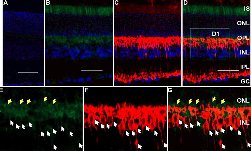
Figure 5. Localization of Cre expression in the rod bipolar cells
Immunohistochemical detection of Cre-activated β-gal expression in the inner nuclear layer of six-week-old SMOPC1 mice. A,B: Representative immunohistochemistry (IHC) staining with a polyclonal anti-β-gal (green) antibody using retinal sections from a wild-type littermate and a SMOPC1/R26R double transgenic mouse, respectively. C: IHC staining of the identical section used in Panel B with a monoclonal anti-protein kinase C (PKC) antibody (red) for rod bipolar cells. D: Merged image of Panels B and C. Blue is DAPI nuclear staining. E-G: Enlarged images (identical area as D1) corresponding to B-D, respectively, without showing DAPI staining. The scale bar represents 50 μm. Photoreceptor inner segment (IS), outer nuclear layer (ONL), outer plexiform layer (OPL), inner nuclear layer (INL), inner plexiform layer (IPL), and ganglion cell layer (GC) are labeled. In addition to the strong β-gal staining in the photoreceptor inner segment, β-gal staining was also observed in the photoreceptor terminals (yellow arrows) and the INL (white arrows). The Cre-activated β-gal in the INL of the SMOPC1 mice was co-localized to the rod bipolar cells (white arrows).